DNA WORLD FOMOUS
Saturday, November 27, 2010
Domain Owner Ship Page
"This post conforms my ownership of the site and that this site adheres to google adsense program policies and terms and conditions"
Major Advance Made On DNA Structure
ScienceDaily (May 4, 2005) — CORVALLIS, Ore. -- Oregon State University researchers have made significant new advances in determining the structure of all possible DNA sequences -- a discovery that in one sense takes up where Watson and Crick left off, after outlining in 1953 the double-helical structure of this biological blueprint for life.
ne of the fundamental problems in biochemistry is to predict the structure of a molecule from its sequence -- this has been referred to as the "Holy Grail" of protein chemistry.
Today, the OSU scientists announced in the Proceedings of the National Academy of Sciences that they have used X-ray crystallography to determine the three-dimensional structures of nearly all the possible sequences of a macromolecule, and thereby create a map of DNA structure.
As work of this type expands, it should be fundamentally important in explaining the actual biological function of genes - in particular, such issues as genetic "expression," DNA mutation and repair, and why some DNA structures are inherently prone to damage and mutation. Understanding DNA structure, the scientists say, is just as necessary as knowing gene sequence. The human genome project, with its detailed explanation of the genetic sequence of the entire human genome, is one side of the coin. The other side is understanding how the three-dimensional structure of different types of DNA are defined by those sequences, and, ultimately, how that defines biological function.
"There can be 400 million nucleotides in a human chromosome, but only about 10 percent of them actually code for genes," said Pui Shing Ho, professor and chair of the OSU Department of Biochemistry and Biophysics. "The other 90 percent of the nucleotides may play different roles, such as regulating gene expression, and they often do that through variations in DNA structure."
"Now, for the first time, we're really starting to see what the genome looks like in three dimensional reality, not just what the sequence of genes is," Ho said. "DNA is much more than just a string of letters, it's an actual structure that we have to explore if we ever hope to understand biological function. This is a significant step forward, a milestone in DNA structural biology."
In the early 1950s, two researchers at Cambridge University -- James Watson and Francis Crick -- made pioneering discoveries by proposing the double-helix structure of DNA, along with another research group in England about the same time. They later received the Nobel Prize for this breakthrough, which has been called the most important biological work of the past century and revolutionized the study of biochemistry. Some of the other early and profoundly important work in protein chemistry was done by Linus Pauling, an OSU alumnus and himself the recipient of two Nobel Prizes.
However, Watson and Crick actually identified only one structure of DNA, called B-DNA, when in fact there are many others -- one of which was discovered and another whose structure was solved at OSU in recent years -- that all have different effects on genetic function.
Aside from the genetic sequence that DNA encodes, the structure of the DNA itself can have profound biological effects, scientists now understand. Until now, there has been no reliable method to identify DNA structure from sequence, and learn more about its effects on biological function.
In their studies, the OSU scientists used X-ray examination of crystalline DNA to reconstruct exactly what the DNA looks like at the atomic level. By determining 63 of the 64 possible DNA sequences, they were able to ultimately determine the physical structure of the underlying DNA for all different types of sequences. Another important part of this study is the finding that the process of DNA crystallization does not distort its structure.
"Essentially, this is a proof of concept, a demonstration that this approach to studying DNA structure will work, and can ultimately be used to help understand biology," Ho said.
For instance, one of the unusual DNA structures called a Holliday junction, whose structure was co-solved at OSU about five years ago, apparently plays a key role in DNA's ability to repair itself -- a vital biological function.
A more fundamental understanding of DNA structure and its relationship to genetic sequences, researchers say, helps set the stage for applied advances in biology, biomedicine, genetic engineering, nanotechnology and other fields.
The recent work was supported by grants from the National Institutes of Health and the National Science Foundatio
DNA structure – Fine-tuning and optimization
Highly repetitive nucleotide sequences lack stability and mutate readily. However, a study involving the genomes of different organisms at the University of California suggests that codon usage in genes is actually designed to avoid the type of repetition that leads to unstable sequences! Further research indicates that codon usage in genes is also set up to maximize the accuracy of protein synthesis at the ribosome.
Furthermore, the components which comprise the nucleotides also appear to have been carefully chosen in view of enhanced performance. Nucleotides that form the strands of DNA are complex molecules which consist of both a phosphate moiety and a nucleobase (adenine, guanine, cytosine or thymine) joined to a five-carbon sugar (deoxyribose). In RNA, the five-carbon sugar ribose replaces deoxyribose.
The phosphate group of one nucleotide links to the deoxyribose unit of another to form the backbone of the DNA strand. The nucleobases form the ‘ladder rungs’ when the two strands align and twist to form the classical double-helix structure.
Scientists have long known that a myriad of sugars and numerous other nucleobases could have conceivably become part of the cell’s information storage medium (DNA). But why do the nucleotide subunits of DNA and RNA consist of those particular components? Phosphates can form bonds with two sugars simultaneously (called phosphodiester bonds) to bridge two nucleotides, while retaining a negative charge. This makes this chemical group perfectly suited to form a stable backbone for the DNA molecule. Other compounds can form bonds between two sugars but are not able to retain a negative charge. The negative charge on the phosphate group imparts the DNA backbone with stability, thus giving it protection from cleavage by reactive water molecules. Furthermore, the intrinsic nature of the phosphodiester bonds is also finely-tuned. For instance, the phophodiester linkage that bridges the ribose sugar of RNA could involve the 5’ OH of one ribose molecule with either the 2’ OH or 3’ OH of the adjacent ribose molecule. RNA exclusively makes use of 5’ to 3’ bonding. As it turns out, the 5’ to 3’ linkages impart far greater stability to the RNA molecule than does the 5’ to 2’ bonds.
Why do deoxyribose and ribose serve as the backbone constituents of DNA and RNA respectively? Both are five-carbon sugars which form five-membered rings. It is possible to make DNA analogues using a wide range of different sugars that contain four, five and six carbons that can form five- and six-membered rings. But these DNA variants possess undesirable properties as compared to DNA and RNA. For instance, some DNA analogues do not form double helices. Others do, but the nucleotide strands either interact too tightly or too weakly, or they display inappropriate selectivity in their associations. Furthermore, DNA analogues made from sugars that form 6-membered rings adopt too many structural conformations. In this event, it becomes exceptionally difficult for the cell’s machinery to properly execute DNA replication and transcription. Other research shows that deoxyribose uniquely provides the necessary space within the backbone region of the double helix of DNA to accommodate the large nucleobases. No other sugar fulfils this requirement.
DNA structure – An overview
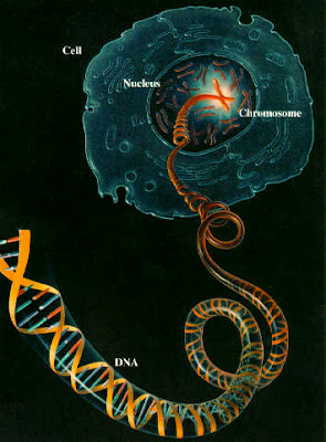
DNA consists of two chainlike molecules (polynucleotides) that twist around each other to form the classic double-helix. The cell’s machinery forms polynucleotide chains by linking together four nucleotides. The nucleotides which are used to build DNA chains are adenosine (A), guanosine (G), cytidine (C), and thymidine (T). DNA houses the information required to make all the polypeptides used by the cell. The sequence of nucleotides in DNA strands (called a ‘gene’) specifies the sequence of amino acids in polypeptide chains.
Clearly a one-to-one relationship cannot exist between the four nucleotides of DNA and the twenty amino acids used to assemble polypeptides. The cell therefore uses groupings of three nucleotides (called ‘codons’) to specify twenty different amino acids. Each codon specifies an amino acid.
Because some codons are redundant, the amino acid sequence for a given polypeptide chain can be specified by several different nucleotide sequences. In fact, research has confirmed that the cell does not randomly make use of redundant codons to specify a particular amino acid in a polypeptide chain. Rather, there appears to be a delicate rationale behind codon usage in genes.
sexual reproduction and meiosis
DNA also replicates reliably in the process of meiosis, which happens before sex cells ( gametes ) are produced, but only half the normal number of chromosomes (and hence genes, and DNA) are distributed to each gamete. The sharing process in halving the number of chromosomes also includes elements of "scrambling" which introduce variation, so each gamete has a unique DNA content.
Meiosis
qmei1 qmei2 qmei3 qmei4 qmei5 qmei6
Firstly, chromosomes associate with
their "partners", then each replicates,
perhaps with exchange of genetic
material. Secondly, chromatids
separate - as in mitosis. qmei7 qmei8 qmei9
In meiosis, the nucleus divides twice to produce 4 nuclei, which then form into 4 genetically different sex cells (gametes), each containing half the number of chromosomes of the original cell (23 in human cells)
Therefore every organism produced as a result of sexual reproduction varies. However, the DNA built into the nucleus of a gamete may also be changed due to a random event called a mutation, which may alter or even prevent the normal activity of a gene inside cells. In this way a different form of the gene, called an allele , is produced, and will possibly be passed on to the next generation. Because each chromosome usually has a partner in the nucleus, the effect of a mutant allele may be hidden by the DNA of a normal allele of that gene which produces a normal characteristic.
DNA and NUCLEAR DIVISION
These 2 double strands form the 2 sections of chromosomes (called chromatids) that are easily seen when a cell is about to divide. In mitosis the chromosomes are then evenly distributed to different ends of the cell, ready to be incorporated into 2 new cells when the cell itself divides.
animation - mitosis
Mitosis
For clarity, only 2 pairs of chromosomes are shown in these diagrams
mit1 mit2 mit3 mit4 mit5
In mitosis, the nucleus divides once to produce 2 nuclei, which then form into 2 genetically identical "ordinary" cells, containing the same number of chromosomes as the original cell (46 in human cells)
Because of the reliability of the replication of DNA and mitosis, offspring resulting from asexual reproduction do not usually vary at all, which is the basis of taking cuttings, etc. Similarly, multicellular organisms consists of a harmonious population of identical cells derived from one initial cell, the fertilised egg or zygote. However, in some cases (about 1 in a million) there may be an error in the copying process; an incorrect copy of the DNA will be passed on to any (body) cells produced following cell division. This may be the cause of different types of cancer, which are associated with exposure to radiation or chemicals and viruses which damage DNA.
How DNA replicates
Understanding this goes a long way to explaining how nuclei divide in the process of mitosis , which results in identical copies of chromosomes being transferred during ordinary cell division.
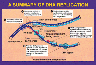
Before a cell divides, its nucleus must divide. But before that happens, the chromosomes must have become double. So the first stage is that DNA which the chromosomes contain must replicate , i.e. become double, by making copies of itself.
The 2 strands of the DNA double helix can separate, under the influence of special enzymes in the nucleus, but each half remains attached along its length, like the 2 sections of a zip, because the sides of the strands are strongly joined.
In the diagrams below, write in the letters for the various bases (using the first few as a key). This should help you understand the results of the process.
Original DNA molecule Unzipping DNA
New bases being added 2 Double DNA strands
DNA replication
Each strand then acts as a basis for rebuilding the missing other strand from which it has been separated. It is said that each strand forms a template on which it reforms its complementary strand.
Enzymes within the nucleus match the appropriate base, which is already attached to strand side subunits, so that A fits against T, G against C, T against A and C against G, according to shape.
Other possibilities are not allowed, so the copying process is accurate in the vast majority of cases.
The result is that one double strand is converted into two identical double strands.
It is interesting to note that each "new" double strand is in fact half composed of a section of the previous DNA molecule, together with a completely new section built up from individual bases.
Subscribe to:
Comments (Atom)


